Swallowing Disorders in Singapore: Causes, Assessment, and Treatment Options
- Vyas Prasad
- May 24
- 9 min read

Updated: May 28
By Dr Vyas M.N. Prasad, FRCS (ORL-HNS) — Consultant Otolaryngologist & Head and Neck Surgeon, Camden Medical Centre, Singapore
Swallowing disorders (dysphagia) occur when the coordinated muscle and nerve action required for swallowing breaks down. In Singapore, common causes include laryngopharyngeal reflux, cricopharyngeal dysfunction, Zenker's diverticulum, neurological conditions such as Parkinson's disease or stroke, and head and neck cancer. Treatment depends on the cause and may include botulinum toxin injection, endoscopic dilation, laser or stapling surgery, or swallowing rehabilitation. Specialist ENT assessment — including flexible nasendoscopy and FEES — is the recommended first step for anyone with persistent swallowing difficulty.
Dysphagia occurs when the coordinated muscle and nerve action of swallowing breaks down
Common causes include LPR, cricopharyngeal dysfunction, Zenker's diverticulum, Parkinson's disease, stroke, and head and neck cancer
Assessment typically involves endoscopy and swallowing studies
Treatment ranges from dietary modification and speech therapy to minimally invasive surgery

Swallowing is one of those bodily functions we perform dozens of times a day without thought — until something goes wrong. When swallowing becomes difficult, painful, or unreliable, the impact on daily life is immediate and often profound. Mealtimes become stressful. Eating in public becomes anxiety-inducing. Weight loss follows. And the fear — sometimes unspoken — that something serious is being missed can dominate every meal.
Difficulty swallowing, medically termed dysphagia, is a symptom with a wide range of causes. Some are straightforward and treatable; others require careful investigation to identify. What they share is that they should not simply be tolerated or attributed to ageing. Swallowing difficulty warrants specialist assessment — and in the majority of cases, there is something meaningful that can be done.
This article explains the anatomy of normal swallowing, the most common causes of swallowing disorders, how they are assessed, and what treatment options are available in Singapore.
How Swallowing Works
Swallowing is far more complex than it appears. It involves the coordinated action of over 30 muscles, controlled by multiple cranial nerves and two swallowing centres in the brainstem. The process unfolds in three phases:
The oral phase — food or liquid is prepared in the mouth, chewed if necessary, and formed into a bolus. The tongue moves the bolus to the back of the mouth and initiates the swallow.
The pharyngeal phase — this is the most complex and critical phase, lasting less than a second. The soft palate rises to close off the nasal passage. The larynx elevates and the epiglottis folds down to cover the airway, directing food into the oesophagus rather than the trachea. The pharyngeal muscles contract in a coordinated wave to propel the bolus downward. The cricopharyngeal muscle — the upper oesophageal sphincter — relaxes to allow passage into the oesophagus.
The oesophageal phase — peristaltic contractions of the oesophageal wall propel the bolus to the stomach.
A failure at any point in this sequence produces dysphagia. The nature of the difficulty — whether it affects liquids, solids, or both; whether it is associated with coughing, regurgitation, or pain; whether it is progressive or intermittent — provides important diagnostic clues.
Symptoms of Swallowing Disorders
Swallowing disorders present in a variety of ways. The most common symptoms include:
Food sticking — a sensation that food catches or lodges in the throat or chest, requiring multiple swallows or sips of water to clear
Coughing or choking during meals — particularly with liquids, which can suggest laryngeal penetration or aspiration
A wet or gurgly voice after eating — indicating food or liquid residue sitting near the larynx
Pain on swallowing (odynophagia) — which may suggest inflammation, infection, or a structural lesion
Regurgitation — food coming back up without the effort of vomiting, sometimes through the nose
Sensation of a lump in the throat — globus, which may be functional or may reflect a structural cause
Recurrent chest infections — silent aspiration, where food or liquid enters the airway without triggering a cough, can cause repeated aspiration pneumonia
Unexplained weight loss — a consequence of reduced oral intake due to dysphagia
Voice change after swallowing — a hoarse or wet voice quality suggesting laryngeal involvement
In infants and children, swallowing difficulties may present as prolonged or tiring feeds, poor weight gain, recurrent respiratory infections, or noisy breathing during feeds.
Any swallowing difficulty that persists for more than two to three weeks, or that is associated with weight loss, voice change, or recurrent chest infections, warrants specialist assessment.
Causes of Swallowing Disorders
Laryngopharyngeal Reflux (LPR)
LPR — the backflow of stomach contents to the throat and larynx — is one of the most common causes of swallowing discomfort seen in ENT practice. The laryngeal and pharyngeal mucosa are exquisitely sensitive to acid and pepsin; even small amounts of reflux cause irritation, swelling, and a sensation of tightness or obstruction. Patients often describe difficulty initiating a swallow, or a persistent globus sensation that makes every swallow feel effortful.
LPR is frequently underdiagnosed because it does not necessarily cause heartburn — the typical symptom of gastro-oesophageal reflux disease. It responds well to dietary modification and acid suppression, though treatment must be sustained for at least eight to twelve weeks to allow the laryngeal mucosa to recover.
Cricopharyngeal Dysfunction
The cricopharyngeus muscle — the upper oesophageal sphincter — must relax precisely during swallowing to allow food to pass from the pharynx into the oesophagus. When this muscle is overactive, fibrotic, or poorly coordinated, it creates a functional obstruction that causes food to stick at the level of the throat.
Cricopharyngeal dysfunction is one of the most treatable causes of dysphagia and is frequently underdiagnosed. Patients describe food sticking high in the throat — not in the chest — and often have residue that pools above the cricopharyngeus and spills over into the larynx, causing coughing or aspiration.
Botulinum toxin injection into the cricopharyngeus — performed endoscopically under general anaesthesia — reduces muscle tone and can produce immediate and dramatic improvement in swallowing. The effect typically lasts three to six months; in many patients, a single or repeat injection is sufficient for long-term benefit as the swallowing mechanism adapts. Where the dysfunction is more severe or structural, cricopharyngeal myotomy — surgical division of the muscle — provides a more permanent solution.
Zenker's Diverticulum
A Zenker's diverticulum is a pouch that forms at the junction of the pharynx and oesophagus — at the point where the cricopharyngeus muscle meets the inferior pharyngeal constrictor, a structurally weak area known as Killian's dehiscence. Food collects in this pouch, causing regurgitation of undigested food (often hours after eating), halitosis, a gurgling sensation in the neck, and progressive dysphagia.
It is more common in older patients and is strongly associated with cricopharyngeal dysfunction — the elevated pressure in the pharynx from a poorly relaxing sphincter drives the formation of the pouch over time.
Treatment is surgical. The minimally invasive approach — endoscopic stapling or laser division of the common wall between the pouch and the oesophagus — is performed through the mouth under general anaesthesia, with no external incision. This has replaced open surgery as the standard approach in most cases and allows a rapid recovery. In selected patients, a flexible endoscopic approach using a diverticuloscope is also available.
Pharyngeal and Oesophageal Strictures
Narrowing of the pharynx or oesophagus — from chronic reflux, previous radiotherapy, caustic ingestion, or surgical scarring — causes progressive dysphagia, initially for solids, later for soft foods, and eventually for liquids. The history is typically one of gradual worsening over months.
Endoscopic dilation — stretching the narrowed segment using progressively sized dilators or a balloon — is the primary treatment and can be performed as a day procedure under sedation or general anaesthesia. Some strictures require repeat dilation; others respond durably to a single session.
Neurological Causes
The swallowing mechanism depends on intact neurological control — the brainstem swallowing centres and multiple cranial nerves (V, VII, IX, X, XII). Neurological conditions that disrupt this control cause oropharyngeal dysphagia, characterised by difficulty initiating the swallow, coughing or choking, and aspiration.
Common neurological causes include:
Stroke — the most common neurological cause of acute dysphagia; most patients recover swallowing function but a proportion require prolonged rehabilitation and occasionally surgical intervention
Parkinson's disease — progressive swallowing difficulty is common and is a significant source of morbidity, often complicated by silent aspiration and pneumonia
Motor neuron disease / ALS — bulbar involvement causes progressive pharyngeal dysphagia; management focuses on maintaining safe oral intake and planning for nutritional support
Multiple sclerosis — dysphagia occurs in a proportion of patients, particularly with brainstem lesions
Head and neck cancer treatment — surgery and radiotherapy to the pharynx and larynx can damage the swallowing mechanism, sometimes causing persistent dysphagia requiring long-term rehabilitation
Management of neurological dysphagia is multidisciplinary — involving the ENT surgeon for assessment and specific interventions, the speech and language therapist for swallowing rehabilitation, and the neurologist or rehabilitation physician for management of the underlying condition.
Head and Neck Cancer
Any malignancy involving the pharynx, larynx, tongue base, or oesophagus can cause progressive dysphagia. New onset dysphagia — particularly when progressive, associated with weight loss, a neck lump, or voice change — should always prompt specialist assessment to exclude a tumour. Assessment includes flexible nasendoscopy and, where indicated, imaging and endoscopic biopsy.
Laryngomalacia and Congenital Causes in Children
In infants, the most common cause of feeding difficulty and noisy breathing is laryngomalacia — a condition in which the supraglottic structures (the tissues above the vocal folds) are abnormally soft and collapse inward during breathing and feeding. Most cases are mild and resolve with growth; severe cases causing significant feeding difficulty, failure to thrive, or oxygen desaturation require surgical treatment — supraglottoplasty, performed endoscopically.
Other congenital causes of swallowing difficulty in infants include cleft palate, tracheo-oesophageal fistula, and vascular rings compressing the oesophagus.
How Swallowing Disorders Are Assessed
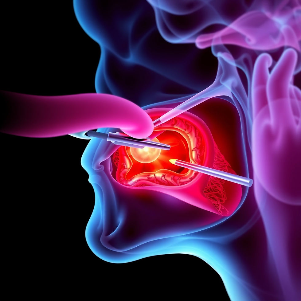
Flexible nasendoscopy
A brief, well-tolerated examination in clinic using a small flexible camera passed through the nose. This allows direct visualisation of the throat, voice box, and the area around the cricopharyngeus, and can identify structural causes of dysphagia, pooling of secretions, and laryngeal abnormalities.
FEES — Fibreoptic Endoscopic Evaluation of Swallowing
FEES is a functional swallowing assessment performed in the clinic. A flexible endoscope is passed through the nose to the level of the larynx, and the patient is given food and drink of different consistencies to swallow while the passage of the bolus through the pharynx is observed directly. FEES identifies penetration (food entering the laryngeal inlet) and aspiration (food passing below the vocal folds into the trachea), and assesses the efficiency of pharyngeal clearance. It is the investigation of choice for oropharyngeal dysphagia.
Videofluoroscopy (modified barium swallow)
A dynamic X-ray study in which the patient swallows barium-coated food and liquid of different consistencies while being filmed. It provides a complementary view to FEES — in particular, it allows assessment of the oral phase and the oesophageal phase, which are not visible on FEES. Performed in collaboration with the radiology department and the speech and language therapist.
Rigid endoscopy under general anaesthesia
Where a structural lesion is suspected — a stricture, a Zenker's diverticulum, or a pharyngeal tumour — rigid endoscopy allows detailed inspection of the pharynx and upper oesophagus and permits biopsy or therapeutic intervention (dilation, myotomy) at the same sitting.
CT and MRI
Imaging is used to assess structural causes of dysphagia — tumours, vascular compression, cervical osteophytes — and is arranged where the clinical assessment suggests a lesion that cannot be fully characterised endoscopically.
Treatment Options
Treatment is tailored to the underlying cause:
LPR — dietary modification, acid suppression, voice therapy
Cricopharyngeal dysfunction — botulinum toxin injection; cricopharyngeal myotomy for refractory cases
Zenker's diverticulum — endoscopic stapling or laser myotomy; open surgery for large or complex pouches
Strictures — endoscopic dilation, repeated as necessary
Neurological dysphagia — swallowing rehabilitation with speech and language therapy; dietary texture modification; feeding tube placement where oral intake cannot be maintained safely
Head and neck cancer — treatment of the primary tumour combined with swallowing rehabilitation; surgical reconstruction of the swallowing pathway where indicated
Laryngomalacia in infants — observation for mild cases; supraglottoplasty for severe cases
When to Seek Assessment
I would recommend specialist assessment for any of the following:
Swallowing difficulty persisting for more than two to three weeks
Progressive difficulty — initially with solids, then soft foods, then liquids
Swallowing associated with coughing, choking, or a wet voice
Recurrent chest infections without a clear cause
Regurgitation of undigested food
Pain on swallowing
Unexplained weight loss
Any new swallowing difficulty in a patient with a history of head and neck cancer or radiotherapy
Swallowing disorders are rarely self-limiting, and the consequences of untreated aspiration — recurrent pneumonia, malnutrition, and reduced quality of life — are significant. Early assessment leads to earlier treatment and better outcomes.
To arrange a consultation with Dr Vyas Prasad at Camden Medical Centre, please contact us. Assessment includes flexible nasendoscopy in clinic and access to the full range of swallowing investigations and treatment options, including botulinum toxin injection, endoscopic dilation, and minimally invasive surgical procedures.


My father had a web in his esophagus which Dr Vyas ballooned and now has normal swallowing. Good treatment option. Thanks.