Head and Neck Cancer in Singapore: Symptoms, Diagnosis, and Treatment
- Vyas Prasad
- May 24
- 9 min read

Updated: May 28
By Dr Vyas M.N. Prasad, FRCS (ORL-HNS) — Consultant Otolaryngologist & Head and Neck Surgeon, Camden Medical Centre, Singapore
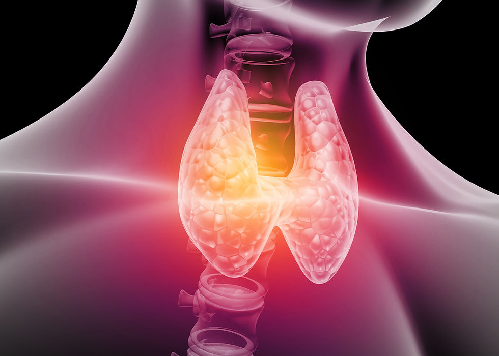
Head and neck cancer covers the throat, voice box, mouth, nose, salivary glands, and neck lymph nodes
Nasopharyngeal cancer (NPC) is particularly prevalent in Singapore, especially in men of Chinese ethnicity
Key warning signs include a persistent neck lump, hoarseness over 3 weeks, a mouth ulcer that won't heal, and unexplained weight loss
Early diagnosis significantly improves treatment outcomes

Head and neck cancer encompasses malignancies of the throat, voice box, mouth, nose, salivary glands, and neck lymph nodes. In Singapore, nasopharyngeal cancer (NPC) is particularly prevalent, especially in men of Chinese ethnicity. Key warning signs include a persistent neck lump, hoarseness lasting more than 3 weeks, a mouth ulcer that won't heal, and one-sided nasal or ear symptoms. Treatment combines surgery, radiotherapy, and chemotherapy depending on cancer type and stage.
Dr Vyas Prasad offers specialist head and neck surgical assessment at Camden Medical Centre, 1 Orchard Boulevard, Singapore.
Head and neck cancer is a term that encompasses a group of malignancies arising from the structures of the upper aerodigestive tract — the mouth, throat, voice box, nasal cavity, sinuses, salivary glands, and the skin of the head and neck. Together, these cancers account for a significant proportion of all cancers diagnosed in Singapore and across Southeast Asia, where certain risk factors — particularly tobacco use, areca nut (betel nut) chewing, and Epstein-Barr virus — are prevalent.
What unites these cancers, despite their anatomical diversity, is the importance of early detection. Many head and neck cancers are highly treatable when diagnosed at an early stage — and many of the symptoms that warrant investigation are ones that patients experience for weeks or months before seeking specialist advice, often attributing them to minor infections or stress.
This article explains the main types of head and neck cancer, the symptoms to look out for, how diagnosis is approached, and what treatment involves.
Types of Head and Neck Cancer
Nasopharyngeal Cancer (NPC)
Nasopharyngeal cancer arises in the nasopharynx — the upper part of the throat, behind the nose. It is of particular significance in Singapore and Southeast Asia, where it is disproportionately common compared to Western populations. The incidence is highest in men of Chinese ethnicity, and Epstein-Barr virus (EBV) infection plays a central role in its development.
NPC often presents at a relatively advanced stage because the nasopharynx is not easily visualised and early symptoms are non-specific. The most common presenting feature is a painless lump in the neck — enlarged lymph nodes — which may be present before any primary throat symptoms develop. Other symptoms include nasal obstruction on one side, blood-stained nasal discharge, a sensation of fullness or blocked ear on one side (caused by Eustachian tube dysfunction from the tumour), and occasionally facial numbness or double vision if the tumour extends to involve the skull base.
EBV serology — blood tests measuring antibodies to EBV — is used in screening and diagnosis. Biopsy of the nasopharynx, performed under nasendoscopy, confirms the diagnosis. MRI of the head and neck and PET-CT are the key staging investigations.
NPC is highly radiosensitive, and radiotherapy — often combined with chemotherapy — is the primary treatment. Surgery plays a limited role in primary treatment but is important in managing residual or recurrent disease.
Oropharyngeal Cancer
Oropharyngeal cancer arises in the oropharynx — the middle part of the throat, including the tonsils, the base of the tongue, the soft palate, and the posterior pharyngeal wall. There are two distinct subtypes with different biology and behaviour:
HPV-associated oropharyngeal cancer — driven by human papillomavirus, particularly HPV-16 — has increased significantly in incidence globally and now accounts for the majority of oropharyngeal cancers in many populations. It tends to affect younger, non-smoking patients, often without the traditional risk factors of tobacco and alcohol. Paradoxically, HPV-associated tumours have a better prognosis than their non-HPV counterparts. The typical presentation is a painless neck lump — metastatic lymph node — often with a relatively small and easily missed primary in the tonsil or tongue base.
Non-HPV oropharyngeal cancer is more strongly associated with tobacco and alcohol use and tends to present at a more advanced stage with pain, difficulty swallowing, or referred ear pain.
Treatment of oropharyngeal cancer combines surgery and radiotherapy, with the approach determined by tumour stage, HPV status, and the patient's overall condition. Robotic surgery — transoral robotic surgery (TORS) — has become an important tool for resecting tumours of the tonsil and tongue base with minimal external incisions.
Oral Cavity Cancer
Oral cavity cancer encompasses cancers of the lip, tongue (anterior two-thirds), floor of mouth, buccal mucosa, gingiva, and hard palate. Squamous cell carcinoma is the predominant histological type. Risk factors include tobacco use in all forms — smoking, chewing tobacco, and betel nut — and heavy alcohol consumption.
Oral cavity cancer is often visible — a persistent ulcer, a white patch (leukoplakia), a red patch (erythroplakia), or an indurated lump in the mouth that does not heal — and yet is frequently dismissed or treated as a minor lesion for months before specialist assessment. Any oral lesion that has not healed within two to three weeks warrants assessment.
Surgery is the primary treatment for most oral cavity cancers, sometimes combined with post-operative radiotherapy. Reconstruction of the oral cavity following tumour resection — using local flaps, regional flaps, or microvascular free flaps — is an integral part of treatment and requires specialist head and neck surgical expertise.
Laryngeal Cancer
Laryngeal cancer arises from the mucosa of the larynx — the voice box. It is strongly associated with tobacco smoking and heavy alcohol use. The most important early symptom is persistent hoarseness — a change in voice quality lasting more than three weeks, which should always prompt laryngoscopy.
Laryngeal cancer is classified by its location within the larynx:
Glottic cancer (involving the vocal folds) — tends to present early with hoarseness, as even small tumours affect voice quality. Early-stage glottic cancer has an excellent prognosis with either radiotherapy or endoscopic laser surgery.
Supraglottic cancer (above the vocal folds) — often presents later, with dysphagia, throat discomfort, or neck nodes, because early lesions may not affect the voice.
Subglottic cancer (below the vocal folds) — uncommon, tends to present late with airway compromise.
Treatment depends on stage: early laryngeal cancer is treated with radiotherapy or transoral laser microsurgery, with excellent voice outcomes. Advanced disease requires total laryngectomy — removal of the larynx — combined with reconstruction of the swallowing pathway and voice rehabilitation.
Hypopharyngeal Cancer
Hypopharyngeal cancer arises in the lower throat, around the entrance to the oesophagus. It is strongly associated with tobacco and alcohol, tends to present late, and has one of the poorer prognoses among head and neck cancers. The typical presentation is progressive dysphagia and a neck lump. Treatment is multimodal — surgery, radiotherapy, and chemotherapy — often in combination.
Salivary Gland Cancer
Salivary gland tumours — which arise most commonly in the parotid gland — are predominantly benign. However, a proportion are malignant, and the distinction requires histological assessment. The most common malignant salivary gland tumours include mucoepidermoid carcinoma, adenoid cystic carcinoma, and acinic cell carcinoma.
A parotid lump is the typical presentation. Facial nerve involvement — weakness of the face — is a sign of malignant infiltration and requires prompt specialist assessment. Treatment is primarily surgical, with post-operative radiotherapy in higher-risk cases.
Skin Cancer of the Head and Neck
The head and neck region — particularly the face and scalp — is one of the most sun-exposed areas of the body and a common site for cutaneous malignancies: basal cell carcinoma, squamous cell carcinoma, and melanoma. Singapore's tropical climate and outdoor lifestyle make this particularly relevant. Surgical excision with appropriate margins is the primary treatment; reconstruction in a cosmetically sensitive area requires surgical expertise and careful planning.
Warning Signs: When to See a Specialist
Many head and neck cancers are diagnosed late because the early symptoms are non-specific and easily attributed to benign causes. The following symptoms — particularly when persistent for more than two to three weeks — warrant specialist assessment:
A lump in the neck that has not resolved — this is the most common presentation of head and neck cancer
Persistent hoarseness or voice change
Difficulty swallowing or a sensation of food sticking
A mouth ulcer or patch that has not healed
Persistent one-sided nasal obstruction or blood-stained nasal discharge
Blocked ear on one side in an adult — particularly with no history of ear disease
Referred ear pain without an obvious ear cause
Facial numbness or weakness
Unexplained weight loss in conjunction with any of the above
None of these symptoms necessarily indicates cancer — most have benign explanations. But each warrants examination and, where appropriate, investigation. Early diagnosis makes a profound difference to both treatment options and outcomes.
How Head and Neck Cancer Is Diagnosed
Clinical assessment
The assessment begins with a thorough history and clinical examination — including examination of the oral cavity, flexible nasendoscopy to assess the nose, nasopharynx, and larynx, and palpation of the neck lymph nodes. This can usually be performed at the first consultation and provides a great deal of diagnostic information.
Imaging
MRI is the preferred modality for assessing the primary tumour — it provides excellent soft tissue detail and delineates the extent of local invasion. CT of the neck and chest assesses lymph node involvement and identifies distant metastases. PET-CT is increasingly used for staging — it identifies metabolically active disease throughout the body and is particularly valuable in identifying the primary tumour when the presentation is a neck node of unknown origin.
Tissue diagnosis
A tissue biopsy is required to confirm the diagnosis. Depending on the site, this may involve:
Fine needle aspiration cytology (FNAC) of a neck node — a simple, well-tolerated clinic procedure
Endoscopic biopsy of the primary tumour under local or general anaesthesia
Tonsillectomy — in cases of suspected HPV-associated oropharyngeal cancer where the primary is not visible, removing the tonsil on the affected side allows histological examination of the full tonsillar tissue
Multidisciplinary team (MDT) review
All patients with confirmed or suspected head and neck cancer are discussed at a multidisciplinary team meeting, bringing together head and neck surgeons, clinical oncologists, radiologists, pathologists, and specialist nurses. The MDT formulates a recommended treatment plan based on the full clinical picture, which is then discussed in detail with the patient.
Treatment of Head and Neck Cancer
Treatment depends on the cancer type, stage, and anatomical site, as well as the patient's overall health and priorities. The three main modalities — surgery, radiotherapy, and systemic therapy — are used alone or in combination.
Surgery
Surgery plays a central role in many head and neck cancers, either as the primary treatment or as part of a combined approach. The goals of surgery are complete removal of the tumour with clear margins, management of the regional lymph nodes (neck dissection), and reconstruction of the resected area to restore form and function.
Reconstruction after extensive head and neck resection has advanced enormously — microvascular free flap reconstruction, in which tissue with its own blood supply is transferred from a distant site (commonly the forearm, thigh, or fibula), allows complex defects to be rebuilt with excellent functional and aesthetic outcomes.
Radiotherapy
Radiotherapy is used as a primary treatment for many head and neck cancers — particularly NPC and early laryngeal cancer — and as adjuvant treatment after surgery where there is a risk of residual disease. Modern intensity-modulated radiotherapy (IMRT) allows precise delivery of radiation to the tumour while minimising dose to surrounding structures, significantly reducing the side effects of treatment compared to older techniques.
Systemic therapy
Chemotherapy — most commonly cisplatin-based — is used concurrently with radiotherapy in many locally advanced head and neck cancers, enhancing the effect of radiation. Targeted therapies (cetuximab) and immunotherapy (pembrolizumab, nivolumab) are increasingly used, particularly in recurrent or metastatic disease where standard treatment has not been effective.
Why Subspecialty Expertise Matters
The head and neck region contains a concentration of critical structures — the carotid arteries, the jugular veins, the facial nerve, the recurrent laryngeal nerve, the hypoglossal nerve, the brachial plexus, and more — that make surgery in this area technically demanding. Outcomes in head and neck cancer are demonstrably better at centres with high surgical volume and subspecialty expertise, and the functional consequences of treatment — for voice, swallowing, and appearance — are profoundly affected by the quality of the surgery and reconstruction.
Dr Vyas Prasad trained in head and neck surgical oncology at the Royal National Throat Nose and Ear Hospital (Gray's Inn Road), Barts and the London and at the Royal Marsden Hospital London, four of the UK's leading head and neck surgical centres.
He brings this fellowship-level expertise from CHU UCL Namur, Belgium as well to his practice at Camden Medical Centre, 1 Orchard Boulevard, Singapore — where he manages both straightforward and complex head and neck conditions, and works within a multidisciplinary framework for patients requiring oncological care.
Taking the Next Step
If you have a persistent neck lump, a voice change, a mouth lesion, or any of the other symptoms described in this article, the most important step is specialist assessment — sooner rather than later.
To arrange a consultation with Dr Vyas Prasad at Camden Medical Centre, please contact us via WhatsApp or through the contact form on this site. Assessment is thorough, unhurried, and supported by on-site flexible nasendoscopy and access to the full range of investigations needed to reach a clear diagnosis.


Had a skin cancer removed from my face by Dr Vyas
Excellent blog
Worth noting
Thank you for the advice
I read the blog after seeing doctor vyas and was grateful for the information